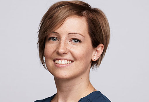
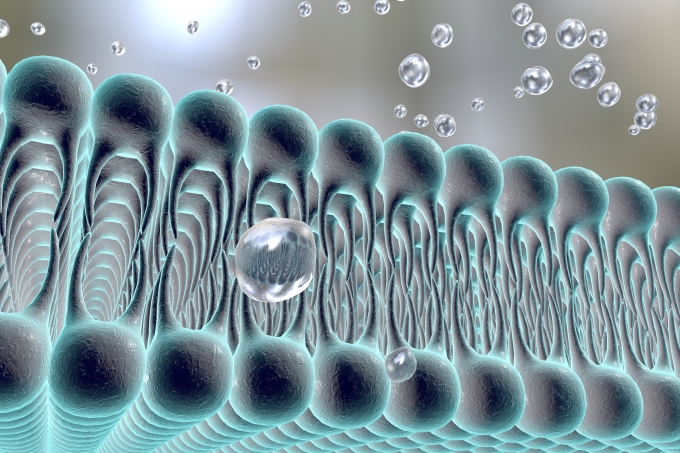
Membrane proteins are large, complex molecules that are part of or interact with cell membranes. They are responsible for regulating processes that help biological cells survive and are the prominent targets for more than fifty percent of prescription drugs on the pharmaceutical market, including drugs that treat neurological disorders and cancers. Despite many advances, membrane proteins cannot be imaged through cryo-electron microscopy at a satisfying resolution, which prevents scientists from gaining a precise understanding of their role in the cell and the associated therapeutic effects of the drugs. Nina Miolane, an assistant professor of electrical and computer engineering at UC Santa Barbara, has received a prestigious National Institutes of Health (NIH) R01 grant to solve the imaging challenge in an effort to answer long-standing questions about membrane proteins and their importance to the cell. The R01 grant program was created to support health-related research and is the oldest grant mechanism provided by the NIH.
“This R01 grant is significant to my research lab, specifically as a new assistant professor,” said Miolane, whose research group, the BioShape Lab, works to solve outstanding challenges in computational biology and medicine. “It is an honor to be chosen by the NIH, and this decision supports our strong belief that geometric learning methods are timely and much needed in biology and medicine where they are deemed to have a very high impact.”
The three-year, $600,000 award will fund Miolane’s project, titled “Improving Membrane Proteins’ 3D Reconstructions with Cryo-Electron Microscopy.” Cryo-electron microscopy (cryo-EM) allows scientists to study macromolecular biological structures and correlate them with their functions in living organisms. Two-dimensional images are generated by firing beams of electrons at proteins that have been frozen in solution, in order to see their biomolecular structure. Three scientists were awarded the 2017 Nobel Prize in Chemistry for developing cryo-EM.
“The focus of the project is to develop a novel mathematical and statistical method that enhances the resolution of cryo-EM reconstruction, particularly targeting membrane proteins,” said Miolane. “We plan to introduce geometric and deep-learning methods to enhance three-dimensional (3D) biological shape reconstruction.”
Geometric learning is an emerging subfield of artificial intelligence that can use the complex geometric information defining shape spaces. Miolane’s research group will use tools from geometry, computer vision, and deep learning to automatically extract, quantify, and analyze crucial shape information from biological datasets. The team plans to assess the performance of its methods through computations and simulations on benchmark datasets of biological images, before testing them against unique datasets of membrane proteins.
“Pushing the field of research in cryo-EM toward our new direction will allow researchers to enhance their understanding of the structure of a wider range of proteins, which are currently very difficult to study,” explains Miolane. “A better understanding of 3D arrangements could lead to improved structure-based drug design for improved selectivity and effectiveness.”

Miolane will collaborate with an array of experts including Frédéric Poitevin, an associate scientist at Stanford University’s SLAC National Accelerator Laboratory; Cornelius Gati, an assistant professor of biology at the University of Southern California; Khanh Dao Duc, an assistant professor of mathematics at the University of British Columbia; and Claire Donnat, an assistant professor of statistics at the University of Chicago.
“The most exciting aspect of this R01 is that it strengthens our collaboration, which in turn allows us to put our ideas into practice and aim for breakthroughs in geometric learning for cryo-electron microscopy,” said Miolane, who added that two terabytes of cryo-EM images are produced at Stanford’s SLAC National Laboratory every day. “This explosion of data, together with our geometric methods, represents an incredible opportunity to advance the frontiers of biomedical knowledge. We’re already discussing how to apply these ideas beyond cryo-EM images.”
The results will also be added to an open-source library called GeomStats, which Miolane and her collaborators launched a few years ago. The platform allows researchers to contribute code involving differential geometry or to use codes without having to delve into complicated mathematical concepts.

Artist depiction of a cell membrane (Katerynakon)
